Click on the video tab to start watching the video.
Lecture marked as incomplete?
In order for the video to be counted as viewed, it must be watched until the very end. If you haven't completed watching 100% of the video and want to ensure that this lecture is recorded on your account, please drag the marker within the video player window to the end of the file. This will allow you to proceed and obtain your certificate.
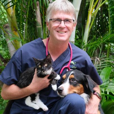
BVSc MVS PG Cert Vet Clin Stud MACVSc (Vet. Emergency and Critical Care; Medicine of Dogs)
Philip graduated from Massey University in New Zealand in 1992, and spent 7 years in small animal practice before undertaking a 3-year residency in veterinary emergency and critical care at the University of Melbourne in 1998.
Following his residency, Philip worked for nearly 6 years at the Animal Emergency Centre in Melbourne, becoming the Senior Veterinarian at the centre in 2004. In 2006, Philip undertook a 1-year surgical externship before moving to Townsville to take up the position of Senior Lecturer in Veterinary Emergency and Critical Care at JCU.
Philip is also co-founder, and director of Vet Education Pty Ltd (www.veteducation.com) – one of Australia’s leading providers of online continuing education for veterinarians and veterinary nurses.
Philip has published numerous manuals and guides concerning emergency medicine, including a CRI manual, haematology and biochemistry interpretation guide, emergency anaesthesia guide, and a ventilation therapy manual for small animals, in addition to being published in peer reviewed literature.
Philip’s key interests in veterinary science include respiratory emergencies, ventilation therapy, envenomations and toxicology.
Login
Accessing this lecture requires a login. Please enter your credentials below!